


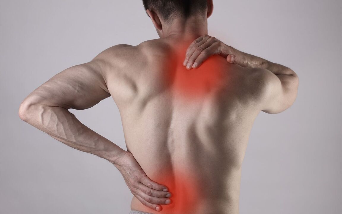
Almost every adult has experienced back pain during his life. This is a very common problem that can be based on various reasons that we will analyze in this article.
Causes of back pain
All causes of back pain can be divided into groups:
Musculoskeletal system:
- Osteochondrosis;
- disc herniation;
- Compression radiculopathy;
- spondylolisthesis;
Inflammatory, including infectious:
- Osteomyelitis
- Tuberculosis
neurological;
Injuries;
Endocrinological;
Vein;
Tumor.
At the first visit to the doctor with back pain, the specialist should determine the cause and type of pain, paying special attention to "red flags" - possible manifestations of potentially dangerous diseases. "Red flags" refer to specific complaints and history data that require a thorough examination of the patient.
"Red Flags":
- age of the patient at the onset of pain: younger than 20 or older than 50;
- a serious spinal cord injury in the past;
- the appearance of pain in patients with cancer, HIV infection or other chronic infectious processes (tuberculosis, syphilis, Lyme disease, etc. );
- temperature;
- weight loss, loss of appetite;
- unusual localization of pain;
- increased pain in a horizontal position (especially at night), weakness in a vertical position;
- No improvement for 1 month or more;
- dysfunction of the pelvic organs, including urinary and defecation disorders, numbness of the perineum, symmetrical weakness of the lower limbs;
- alcoholism;
- drug use, especially intravenous;
- treatment with corticosteroids and/or cytostatics;
- with neck pain, the pulsating nature of the pain.
The presence of one or more symptoms in itself does not mean the presence of a dangerous pathology, but requires the attention and diagnosis of a doctor.
According to the duration of back pain, it is divided into the following forms:
- sharp- pain lasting less than 4 weeks;
- semi-sharp- pain lasting from 4 to 12 weeks;
- chronic- pain lasting 12 weeks or more;
- recurrence of pain- recovery of pain if it did not occur during the last 6 months or more;
- exacerbation of chronic painRecurrence of pain less than 6 months after the previous episode.
Diseases
Let's talk more about the most common musculoskeletal causes of back pain.
Osteochondrosis
This is a disease of the spine, which is based on the erosion of the spinal discs and subsequently the vertebrae themselves.
Is osteochondrosis a pseudodiagnosis? - No. This diagnosis is available in the International Classification of Diseases ICD-10. Currently, doctors are divided into two camps: some believe that such a diagnosis is wrong, while others, on the contrary, often diagnose osteochondrosis. This situation arose because foreign doctors understood osteochondrosis as a disease of the spine related to growth in children and adolescents. However, this term refers to degenerative disease of the spine in people of all ages. Also, frequently established diagnoses are dorsopathy and dorsalgia.
- Dorsopathy is a pathology of the spine;
- Dorsalgia is a benign, non-specific back pain that radiates from the lower cervical vertebrae to the sacrum and can also be caused by damage to other organs.
The spine has several divisions: cervical, thoracic, lumbar, sacral and coccygeal. Pain can occur in any of these areas, which is described by the following medical terms:
- Cervicalgia is pain in the lumbar region of the neck. The intervertebral discs of the cervical region have anatomical features (there are no intervertebral discs in the upper part, and in other parts there is a weakly expressed nucleus pulposus with an average regression of up to 30 years), which makes them more sensitive to stress. and injury that causes stretching of ligaments and early development of degenerative changes;
- Thoracalgia - pain in the thoracic spine;
- Lumbodynia - pain in the lower back (lower back);
- Lumboischialgia is pain in the lower back that radiates to the leg.
Factors leading to the development of osteochondrosis:
- heavy physical labor, lifting and carrying heavy loads;
- low physical activity;
- long sedentary work;
- staying in an uncomfortable position for a long time;
- working for a long time at the computer with a non-optimal monitor location that creates a load on the neck;
- violation of posture;
- congenital structural features and anomalies of the spine;
- weakness of back muscles;
- high growth;
- excess body weight;
- diseases of the joints of the legs (gonarthrosis, coxarthrosis, etc. ), flat feet, club feet, etc. ;
- natural wear with age;
- cigarettes.
disc herniationis the protrusion of the core of the intervertebral disc. It may be asymptomatic or may cause compression of surrounding structures and manifest as radicular syndrome.
Symptoms:
- impaired range of motion;
- feeling of stiffness;
- muscle tension;
- radiating pain to other areas: arms, shoulder blade, legs, groin, rectum, etc.
- "shoots" of pain;
- numbness;
- creeping sensation;
- muscle weakness;
- pelvic disorders.
The localization of pain depends on the level of the hernia.
Disc herniations often resolve on their own within an average of 4-8 weeks.
Compression radiculopathy
Radicular (radicular) syndrome is a complex of manifestations resulting from compression of the spinal roots at the exit points from the spinal cord.
Symptoms depend on the level at which spinal cord compression occurs. Possible manifestations:
- pain in the extremities of a shooting nature with radiation to the fingers, aggravated by movement or coughing;
- numbness or the sensation of flies crawling in a certain area (dermatomes);
- muscle weakness;
- back muscle spasm;
- violation of the power of reflexes;
- positive symptoms of tension (appearance of pain with passive flexion of limbs)
- limitation of spinal mobility.
Spondylolisthesis
Spondylolisthesis is the displacement of the upper vertebra relative to the lower one.
This condition can occur in both children and adults. Women are more affected.
Spondylolisthesis may cause no symptoms with minor displacement and may be an incidental X-ray finding.
Possible symptoms:
- feeling of discomfort
- pain in the back and lower extremities after physical work,
- weakness in the legs
- radicular syndrome,
- pain and reduced sensitivity to touch.
Progression of spinal displacement can lead to spinal stenosis: the anatomical structures of the spine are distorted and enlarged, which gradually causes compression of nerves and blood vessels in the spinal canal. Symptoms:
- constant pain (both at rest and on movement),
- in some cases, the pain may decrease in the lying position,
- pain does not increase with coughing and sneezing;
- the nature of the pain is very strong without withdrawal,
- pelvic organ dysfunction.
With a strong displacement, compression of the vessels can occur, as a result of which the blood supply to the spinal cord is disturbed. This manifests itself with a sharp weakness in the legs, a person can fall.
Diagnostics
Collection of complaintshelps the doctor to suspect the possible causes of the disease, to determine the localization of the pain.
Assessment of Pain Intensity- a very important stage of diagnosis, which allows to choose treatment and evaluate its effectiveness over time. In practice, a Visual Analogue Scale (VAS) is used, which is convenient for the patient and the doctor. In this case, the patient rates the intensity of the pain on a scale from 0 to 10, where 0 is no pain and 10 is the worst pain imaginable.
The interviewallows to determine the factors that cause pain and cause the destruction of the anatomical structures of the spine, to determine the movements and postures that cause, intensify and eliminate pain.
Physical examination:assessment of the presence of spasm of the back muscles, determination of the development of the muscle skeleton, exclusion of the presence of signs of an infectious lesion.
Assessment of neurological status:muscle strength and its symmetry, reflexes, sensitivity.
March Test:performed in cases of suspected lumbar stenosis.
It is important!Patients without "red flags" with a classic clinical picture are not recommended to undergo additional studies.
X-ray:performed with functional tests for suspected instability in the structures of the spine. However, this diagnostic method is non-informative and is mainly implemented with limited financial resources.
Computed tomography (CT) and/or magnetic resonance imaging (MRI):the doctor will prescribe based on clinical data, because these methods have different indications and benefits.
CT |
MRI |
|---|---|
|
|
It is important!In the absence of complaints in most people, degenerative changes in the spine are detected by instrumental examination methods.
Bone densitometry:performed to evaluate bone density (confirmation or exclusion of osteoporosis). This study is recommended for postmenopausal women with a high risk of fracture and always regardless of risk, men over 65 years of age, men over 70 years of age, patients with fractures with minimal history of trauma, long-term use of glucocorticosteroids. The 10-year fracture risk is assessed using the FRAX scale.
Bone scintigraphy, PET-CT:It is carried out in case of suspicion of oncological disease according to other examination methods.
back pain treatment
For acute pain:
- pain relievers are prescribed in a course, mainly from the group of non-steroidal anti-inflammatory drugs (NSAIDs). The specific drug and dosage is selected depending on the severity of the pain;
- maintaining moderate physical activity, special exercises to relieve pain;
It is important!Physical inactivity with low back pain increases the pain, prolongs the duration of symptoms, and increases the likelihood of chronic pain.
- muscle relaxant for muscle spasm;
- it is possible to use vitamins, but according to various studies their effectiveness remains uncertain;
- manual therapy;
- lifestyle analysis and elimination of risk factors.
For subacute or chronic pain:
- the use of painkillers on demand;
- special physical exercises;
- assessment of psychological status, as this can be an important factor in the development of chronic pain and psychotherapy;
- drugs from the group of antidepressants or antiepileptic drugs for the treatment of chronic pain;
- manual therapy;
- lifestyle analysis and elimination of risk factors.
Blockades (epidural injection) or intraosseous blocks are used in radicular syndrome.
Surgical treatment is indicated by the rapid increase in symptoms, the presence of spinal cord compression, significant stenosis of the spinal canal, and ineffectiveness of conservative therapy. Urgent surgical treatment is performed in the following cases: pelvic disorders with numbness in the anogenital region and ascending weakness of the legs (cauda equina syndrome).
Rehabilitation
Rehabilitation should begin as soon as possible and achieve the following goals:
- improving the quality of life;
- removal of pain and if it cannot be completely removed - relief;
- resumption of activity;
- rehabilitation;
- self-service and safe driving training.
Basic rules of rehabilitation:
- the patient should feel responsible for his own health and follow the recommendations, but the doctor should choose the treatment and rehabilitation methods to which the patient can adapt;
- to observe safety rules while performing systematic training and exercises;
- pain is not an obstacle to exercise;
- a reliable relationship must be established between the patient and the doctor;
- the patient should not concentrate and pay attention to the cause of pain in the form of structural changes in the spine;
- the patient should feel comfortable and safe while performing movements;
- the patient should feel the positive effect of rehabilitation on his condition;
- the patient must develop pain response skills;
- the patient must associate the action with positive thoughts.
Rehabilitation methods:
- Walking;
- Physical exercises, gymnastics, gymnastics programs at work;
- Individual orthopedic devices;
- Cognitive Behavioral Therapy;
- Patient Education:
- Avoid excessive physical activity;
- Combating low physical activity;
- Exclusion of long-term static loads (standing, being in an uncomfortable position, etc. );
- avoid hypothermia;
- Sleep organization.
Prevention
Optimal physical activity: strengthens the muscle frame, prevents bone resorption, improves mood and reduces the risk of cardiovascular accidents. The most optimal physical activity is walking more than 90 minutes a week (at least 30 minutes each time, 3 days a week).
With long-term sedentary work, it is necessary to take breaks for warming up every 15-20 minutes and observe the rules of sitting.
Life trick:
how to sit
- avoid overly soft furniture;
- feet should rest on the place obtained by the height of the chair equal to the length of the lower leg;
- it is necessary to sit up to 2/3 of the length of the hips;
- sit straight, maintain the correct posture, the back should sit firmly on the back of the chair in order not to strain the back muscles;
- when reading a book or working on a computer, the head should be in a physiological position (look straight ahead, not always down). For this, it is recommended to use special stands and install the computer monitor at an optimal height.
With long-standing work, it is necessary to change the position every 10-15 minutes, alternately change the supporting leg and, if possible, walk and move in place.
Avoid lying down for long periods of time.
Life trick:
how to sleep
- sleep better on a semi-hard surface. If possible, you can choose an orthopedic mattress so that the spine maintains the physiological curves;
- the pillow should be soft enough and of medium height to avoid stress on the neck;
- it is advisable to put a small pillow under the stomach when sleeping in a prone position.
Quit smoking: If you're having trouble, see your doctor who can refer you to a smoking cessation program.
Frequently asked questions
I use ointments with glucocorticosteroids. Am I at risk of osteochondrosis or osteoporosis? Nope. External glucocorticosteroids (ointments, creams, gels) do not penetrate into the systemic circulation in significant quantities and therefore do not increase the risk of developing these diseases.
Is surgery necessary for every herniated disc? Nope. Surgical treatment is performed only if there is an indication. On average, only 10-15% of patients need surgery.
Should you stop exercising if you have back pain? Nope. If, as a result of additional examination methods, the doctor does not find anything that significantly limits the degree of load on the spine, then you can continue to play sports, but only after undergoing a course of treatment and adding certain exercises. physiotherapy exercises and a swimming course.
If I have a herniated disc, can the back pain go away forever? After a productive course of conservative therapy, they can continue to follow the recommendations of the attending neurologist, follow preventive measures, do regular exercise therapy and swimming.